Episode Transcript
[00:00:11] Speaker A: In the hush of bright screens, tiny filaments.
[00:00:20] Speaker B: Welcome to Base by Bass, the papercast that brings genomics to you wherever you are. Thanks for listening and don't forget to follow and rate us in your podcast app.
[00:00:28] Speaker C: Always great to be back.
[00:00:30] Speaker B: So I want to start today by putting you in a very specific headspace. It's a scenario that plays out in clinics constantly and frankly, it's heartbreaking.
Just imagine you're a parent.
You Notice, say, your 9 year old is stumbling a lot when the sun goes down.
They can't find their seat in a dark movie theater.
[00:00:53] Speaker C: The classic presentation, nyctilopia.
[00:00:55] Speaker B: Right. Night blindness. So you take them to the specialist and they run the tests. They map out the visual field and they give you a name. Retinitis pigmentosa. It's a progressive diagnosis. The vision is going to constrict, basically, like a tunnel closing in. But then comes the next step. You want to know why.
[00:01:10] Speaker C: Naturally, you want the cause. I mean, you want to know the prognosis.
[00:01:13] Speaker B: Exactly. So you sign up for the genetic testing, you give the blood sample, you wait months, and you think, you know, we sequenced 3 billion letters of DNA, surely the answer's in there. And then, yes, silence. The report comes back saying negative.
Or the even more frustrating variant of
[00:01:32] Speaker C: uncertain significance, the dreaded vus.
It's a massive issue in the field. I mean, we are technically capable of reading the entire genome. Yet for inherited retinal diseases, or IRDS, somewhere between 40 and 50% of patients still. Still walk away. Without a molecular diagnosis, that number is wild to me.
[00:01:49] Speaker B: Nearly half.
[00:01:50] Speaker C: Nearly half. Yeah. It's what we call the missing heritability.
We know it's genetic. We can clearly see it tracking through families, but we just. We can't find the typo.
[00:02:00] Speaker B: So that is the mission for this deep dive.
[00:02:02] Speaker C: Yeah.
[00:02:03] Speaker B: We are going hunting for those missing answers. And we're asking, what if the answer isn't actually missing at all? What if it's just hiding in the dark matter of the genome that most standard tests just ignore?
[00:02:13] Speaker C: And perhaps even more intriguingly, what happens when a gene we thought we knew, a gene we thought belonged firmly to the brain, turns out to have a secret life in the eye?
[00:02:22] Speaker B: Yes. Today we celebrate the work of Lynn et al, and a really massive collaborative team. We're talking researchers from the UCL Institute of Ophthalmology, Moorfields Eye Hospital, and the Institute of Molecular and Clinical Ophthalmology. Basel.
[00:02:37] Speaker C: Heavy hitters in the field, for sure.
[00:02:38] Speaker B: Absolutely.
Their paper, which was published on March 5, 2026 in the American Journal of Human Genetics. It effectively solves one of these cold cases.
It's just a masterclass in how to look where others haven't.
[00:02:51] Speaker C: It really is. It highlights that the low hanging fruit in genetics has basically been picked. If you want to solve that remaining 50%, you have to climb higher and you have to dig deeper.
[00:03:00] Speaker B: Right, but before we get into the actual detective work, let's just level set on the condition we mentioned. Retinitis pigmentosa, or rp.
Biologically, what is breaking down here?
[00:03:11] Speaker C: So, RPE is the most common form of inherited retinal disease. It affects about 1 in 3500 people.
Globally. IRDs affect around 5 1/2 million people.
[00:03:21] Speaker B: Wow.
[00:03:22] Speaker C: Yeah, it's significant. You have to picture the retina at the back of the eye like a digital camera sensor. It's lined with photoreceptors, the rods and cones. Exactly. In rp, usually the rods die off first. Those are your low light sensors, hence the night blindness we mentioned earlier. And as they degenerate, the overall structural integrity of the retina starts to fail.
Eventually, the cones, which give you your color and sharp central vision, they die too. And that's when you go blind.
[00:03:50] Speaker B: And the unmet need here isn't just about academic curiosity. I mean, if you don't know the gene, you're just stuck.
[00:03:56] Speaker C: You are totally stuck. You can't enter clinical trials, you can't access the new wave of gene therapies. Well, those are gene specific. If I don't know what to fix and I can't give you the tool to fix it, plus you can't even give the patient an accurate timeline. Some forms of RP are slow, others are rapid. Without the gene, you're really just guessing.
[00:04:14] Speaker B: Okay, so enter the suspect, a gene called FSD1L. Now, I look at genomic papers all day, and I'll be honest, this one was not on my radar for eye disease.
[00:04:22] Speaker C: No, and it shouldn't have been. That's the twist. FSD1L stands for get ready for this. Fibronectin type 3 and spry domain containing 1 like.
[00:04:31] Speaker B: Oh, very catchy.
[00:04:32] Speaker C: Rolls right off the tongue, doesn't it? But until this paper, literally nobody thought it was an eye gene. Its cousin, FSV1, is a known microtubule gene associated with the brain. So finding FSD1L as a definitive cause for blindness is a huge paradigm shift.
[00:04:47] Speaker B: So how did they catch it? If you have 3 billion letters of DNA and you're looking for a typo in a gene, nobody Even suspects. Where do you start?
[00:04:57] Speaker C: They started with a very exclusive club. A cohort of just six individuals from four unrelated families. We had families from the uk, the US and Switzerland.
[00:05:06] Speaker B: Just six people. That sounds incredibly small for a study like this.
[00:05:09] Speaker C: It is small, but that just speaks to the sheer rarity of this mutation. To find them, they utilized massive data sets like the UK 100,000 Genomes Project, just to filter through millions of variants. They were hunting specifically for ultra rare biolelic variants.
[00:05:25] Speaker B: Let's pause on biolelic for a second. We're talking about recessive traits here, right?
[00:05:28] Speaker C: Correct. You need two broken copies, one from mom, one from dad, to get the disease. If you have one working copy, you're a carrier, but your vision is fine. These six patients had mutations in both copies.
[00:05:39] Speaker B: But simply finding a mutation isn't enough to prove anything. I mean, everyone walking around has mutations. How did they prove this specific gene was actually doing something in the eye?
[00:05:49] Speaker C: That is where the deep dive methodology comes in. They couldn't just rely on reading the DNA sequence, they had to look at the context where it operates. So they used single cell RNA sequencing,
[00:05:58] Speaker B: which basically lets them eavesdrop on individual cells to see what genes they are actively using.
[00:06:04] Speaker C: Exactly. They compared human retinal data with mouse data, and they found something really fascinating. This gene, FSD1L, was being expressed. It was being turned on specifically in the photoreceptors.
[00:06:17] Speaker B: And not just any photoreceptors.
[00:06:19] Speaker C: Right. It was highly expressed in the cones and to a slightly lesser extent in the rods, which is super interesting because as we said, RP is usually a rod first disease. It suggests the biology is way more complex than we originally thought, perhaps involving the interplay between how these cells structurally support each other.
[00:06:36] Speaker B: Okay, so the gene is in the crime scene, it's in the eye, but what is it actually doing there?
They use a technique called uxm, or ultrastructure expansion Microscopy. This completely blew my mind when I read it.
[00:06:48] Speaker C: Oh, UXM is brilliant. It's so cool. One of the biggest problems in cell biology is simply that cells are tiny. The machinery inside them is even smaller, actually smaller than the wavelength of light. You just can't see it clearly with a normal light microscope. It's just blurry, it's highly pixelated. Essentially, UXSM solves this by physically expanding the sample itself. Imagine drawing a highly detailed picture on a deflated balloon. It's tiny, it's cramped, you can't make out the lines. Now blow the balloon up, the picture stretches and Suddenly you can see every single detail.
[00:07:21] Speaker B: So they literally inflate the eye tissue?
[00:07:23] Speaker C: In a way, yes. They use a polymer gel to physically expand the tissue about four times its normal size. This let them take super high resolution photos to see exactly where the FSD1L protein was sitting inside the cell.
[00:07:35] Speaker B: And where was it?
[00:07:37] Speaker C: It was sitting right on the connecting cilium and the microtubule axonome.
[00:07:41] Speaker B: Okay, let's demystify those terms for a second. Connecting cilium.
[00:07:44] Speaker C: Right. Think of a photoreceptor like a factory. The main body of the cell makes all the energy in the proteins. And but the antenna, the outer part that actually catches the light, is physically separated from the main body. The connecting cilium is the bridge between them.
[00:07:58] Speaker B: It's like the logistics highway.
[00:07:59] Speaker C: Precisely. It's a very narrow bridge where massive amounts of cargo have to travel back and forth every single second to keep your vision working. And the microtubule axonum is basically the steel skeleton inside that bridge, holding the whole thing up.
[00:08:13] Speaker B: So if FSD1L is broken, the bridge
[00:08:15] Speaker C: becomes unstable, the crucial cargo can't get to the antenna, the antenna starves and the cell dies.
That is the fundamental mechanism of the blindness. Here it's a structural failure of that bridge.
[00:08:27] Speaker B: That makes so much sense. But here is where the story gets really complicated and frankly, really elegant. Because they didn't just find one uniform type of patient.
[00:08:37] Speaker C: No. And this was the puzzle that drove them crazy. In family A, the patients had retinitis pigmentosa, but they also had mild learning disabilities and something called spastic diplegia, which is stiffness in the legs.
[00:08:51] Speaker B: It was a neuroeye syndrome.
[00:08:52] Speaker C: Right. Neurological involvement. But then you look at family D. They had the severe blindness, but absolutely zero neurological issues. No learning disability, no motor issues, just the eye disease.
[00:09:04] Speaker B: How is that even possible? If it's the exact same gene causing it, shouldn't it cause the exact same disease?
[00:09:11] Speaker C: That is the big question. And the answer lies in a really crucial concept called isoforms.
[00:09:16] Speaker B: I love this concept. It's the idea that a gene isn't just one single static instruction.
[00:09:20] Speaker C: Right? Exactly. We often teach genetics in school. Like one gene equals one protein, but it's actually more like one gene equals a recipe book. Depending on which cellular kitchen you're cooking in, you might skip a page or add an extra ingredient. Those different modified versions are called isoforms.
[00:09:35] Speaker B: So the brain uses one recipe from the FSD1L book, and the eye uses a totally different one.
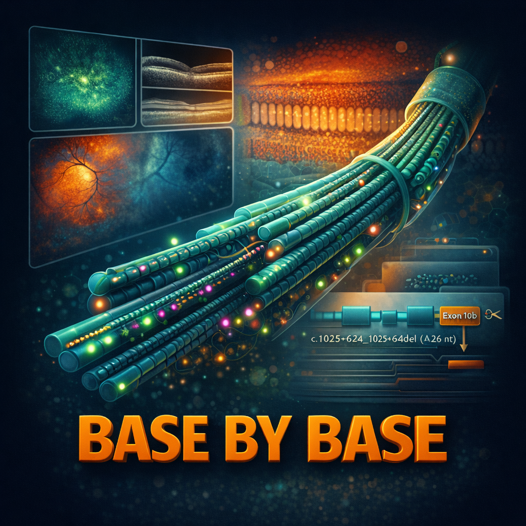
[00:09:39] Speaker C: You hit the nail on the head. The researchers discovered a specific Retina enriched isoformation. The eyes recipe includes a very specific, unique chunk of genetic code called EXON10B.
[00:09:51] Speaker B: EXON10B.
The secret ingredient.
[00:09:54] Speaker C: The secret ingredient that only the eye seems to crave. Now here is the real Sherlock Holmes moment in family D, the family with only blindness. Where was their mutation located?
[00:10:05] Speaker B: It wasn't in the main coding part, was it?
[00:10:07] Speaker C: No, it wasn't. It was a deep intronic deletion. It was buried in the intron, which is the part of the DNA we historically used to call junk DNA or just spacer material.
[00:10:16] Speaker B: The dark matter of the genome.
[00:10:18] Speaker C: Exactly. But this specific patch of dark matter contained the vital instructions for the cellular splicing machinery to say, hey, make sure you include exon10b. Because that specific instruction was deleted, the cell couldn't make the eye recipe.
The brain recipe doesn't even use Exon 10B. So the brain recipe was spliced together perfectly fine. The brain was completely spared.
[00:10:39] Speaker B: That is just incredibly elegant. So by that logic, in family A. Weird. Where they had both the brain and the eye issues, I assume their mutation broke something common to both recipes.
[00:10:49] Speaker C: Spot on. Family A had mutations like frameshifts or missense mutations that wrecked the core protein structure regardless of which isoform was being built. So both the brain and the eye systems failed.
[00:11:00] Speaker B: This completely redefines how we look at the disease. It's not just a binary Is the gene broken or not? It's which version of the gene is broken and in which specific tissue.
[00:11:11] Speaker C: It turns the disease into a spectrum. It perfectly explains why clinical presentations can vary so wildly from patient to patient. And to actually verify this mechanism, they use something called minogene assays.
[00:11:23] Speaker B: Which sounds kind of adorable, but I assume it's highly technical.
[00:11:26] Speaker C: It's a very powerful tool. Since the whole gene is huge, they can't easily test the entire thing in a petri dish. So they literally build a mini version containing just the relevant exons and introns. They put that synthetic gene into cells, specifically ARPE19 cells, which are retinal pigment epithelial cells. And they just watched how the splicing machinery handled the code.
[00:11:48] Speaker B: Kind of like running a spell check simulation on the DNA.
[00:11:50] Speaker C: Yes, and it confirmed that the deep intronic mutation in family D specifically caused the machinery to skip Exon 10B. It proved the mechanism definitively. It wasn't just a hypothesis. They literally watched the cell fail to read the eye recipe.
[00:12:06] Speaker B: Wow.
So, zooming out for a second. We have a new gene, FSD1L. We know it lives on the Connection bridge of the photoreceptor. We know it has a special eye only mode involving exon10b.
Why does this matter to the listener today or to the doctor seeing a patient tomorrow?
[00:12:22] Speaker C: The immediate impact is totally diagnostic. As we said, we have thousands of people in that 40 to 50% unsolved category. We need to start screening them for FSD1L mutations immediately.
[00:12:33] Speaker B: That could literally end the diagnostic odyssey for a lot of families right there, immediately.
[00:12:37] Speaker C: Yes, but the much bigger lesson here is about how we fundamentally do genetic testing. Standard whole exome sequencing, which is the absolute workhorse of clinical genetics right now, only looks at the exons, the coding parts.
[00:12:49] Speaker B: So it would miss family D entirely.
[00:12:51] Speaker C: It would miss them 100% of the time.
This paper is a massive wake up call that we need to be looking at the introns, we need whole genome sequencing, and more importantly, we need to know what we are looking for. When we stare into those non coding regions.
[00:13:06] Speaker B: It's like searching for your keys, right? If you only look under the street lamp because the light is better, you're going to miss them if you drop them out in the shadows. Exome sequencing is just the street lamp.
[00:13:17] Speaker C: That's a perfect analogy. And biologically, this connects two totally different medical fields.
You have neurologists seeing kids with learning delays and ophthalmologists seeing kids with blindness.
This gene proves they need to be talking to each other. The symptoms are connected at the root. It's what we call a syndromic versus non syndromic presentation. But the root cause is the exact same gene, just sliced differently by the cell.
[00:13:43] Speaker B: What about treatment though? Does knowing it's a transport bridge problem help us actually fix it?
[00:13:49] Speaker C: It's the essential first step. You can't fix a car if you don't know which part is smoking. Now we know it's a failure of the ciliar transport. Researchers can start looking at drugs that stabilize microtubules, or looking at gene therapy. If we want to treat family D, we need to deliver a healthy copy of the gene to the retina.
[00:14:08] Speaker B: But we better make absolutely sure we deliver the version with Exon 10B.
[00:14:12] Speaker C: Exactly. If you just deliver the standard brain version to the eye, it might might not work at all. Or worse, it might interfere with the remaining function. The details matter. The isoforms matter. This level of specificity is crucial for the next generation of genetic medicine.
[00:14:28] Speaker B: It really feels like we are just scratching the surface of the dark genome.
Every time we think we've mapped it out, we find a trapdoor leading to a Whole new room.
[00:14:37] Speaker C: The genome is definitely not a static document. It's incredibly dynamic. It's a fluid library of recipes that changes based on whether you're in the heart, the liver or the eye. And we are just now learning how to read. The marginali are the tiny notes in the margins that tell the cell how to actually read the book.
[00:14:52] Speaker B: So to bring this all together, what's the core takeaway for today's deep dive?
[00:14:57] Speaker C: I'd say it's twofold. We've identified FSD1L as a significant new cause of blindness. It was hiding in plain sight because we weren't looking at the right isoforms, specifically the retina enriched version containing Exon 10B. And we've proven that looking at the junk DNA, the introns, can finally explain why a disease hits one organ but completely spares another.
[00:15:18] Speaker B: And biologically, we've reinforced that. The photoreceptor connecting cilium is a critical chokepoint for vision. If that bridge fails, sight fails.
[00:15:26] Speaker C: It's a tragedy for the cell, certainly, but a real triumph for science that we finally understand how it happens.
[00:15:32] Speaker B: It solves the mystery for these specific families, which is amazing, but I feel like it opens up a much larger, almost philosophical question for the field.
[00:15:40] Speaker C: It really does. And this is what I want you, the listener, to really just chew on after this.
[00:15:44] Speaker B: Go for it.
[00:15:44] Speaker C: We know half of these retinal cases are unsolved. We just found one answer hiding in a tissue specific isoform.
So what does this mean for all the other conditions out there? The heart conditions, the kidney failures, the liver diseases that are currently labeled idiopathic or unknown cause?
[00:16:02] Speaker B: Are they really unknown or are we
[00:16:04] Speaker C: just reading the wrong recipe? Are the answers for millions of patients hiding in alternative isoforms that we haven't mapped yet simply because we're only looking at the standard canonical version of the gene?
[00:16:15] Speaker B: That is a very provocative thought. The idea that the cure, or at least the cause, is sitting right there in the data we already possess, but we just haven't learned the specific dialect of that tissue yet.
[00:16:26] Speaker C: It's the next frontier. We absolutely have to stop looking at the genome as a one size fits all blueprint. It is bespoke. It is tailored to every tissue, and our diagnostics need to be just as tailored if we want to solve that remaining 50%.
[00:16:38] Speaker B: Well, on that note, we have reached the end of our deep dive into the dark corners of the genome.
This episode was based on an Open Access article. Under the CC BY 4.0 license, you can find a direct link to the paper and the license in our episode description. If you enjoyed this, follow or subscribe in your podcast app and leave a five star rating. If you'd like to support our work, use the donation link in the description now. Stay with us for an original track created especially for this episode and inspired by the article you've just heard about.
Thanks for listening and join us next time as we explore more science base by base.
[00:17:21] Speaker A: In the hush of bright screens tiny filaments align Silver threads that carry every scattered sign A name like a whisper fst one along the line Tracing light through narrow halls where shadow meets design.
There was a gap in a pattern, a slit the eye can feel an X unfolded out of sight the traffic made real when the map forgets the doorway Signals drift and peace.
Follow the light along the axle don't let it fade we hold the missing pieces up until the dark unwinds from shadow to a filament that finds itself way home we learn the language of the eye and teach the light to roam.
Six faces in the pedigrees of spectrum in their gaze from steady dust to restless nights the redneck keeps its ways as it forms like different passports Some lost at the gate A quiet splice, a hidden cut let changes fade.
And a splice A tiny deletion accent and be raised Cells reroute their luggage Outer segments left unlaced but knowing where the pathway breaks is where the hope is TRA.
So follow the light along the ax suddenly.
Restored A signal strong enough to sing Bring those sticks on back to the road Let every photons fall spring.